200mm focal length double monochromator / spectrometer, McPherson Model 275D

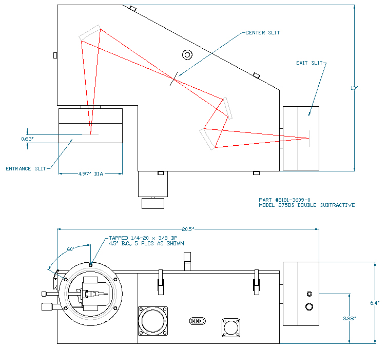
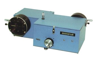
Model 275D Double Monochromator

The Model 275D is a dedicated double grating monochromator providing additive or subtractive dispersion. The 275D compact housing contains the scanning mechanism and mechanical linkage of one precision wavelength sine drive for two gratings. Wavelength control is via the 789A-3 digital scan control with a single motor.The optically and mechanically coupled modified Seya-Namioka systems are equipped with aberration corrected concave holographic gratings. Spectrally agile, the model 275D features all first surface optics for the best UV-Vis-NIR response. Double monochromators are used for a variety of applications that require extremely low levels of scattered or stray light.

Model 275D PDF Data Sheet


Specifications & Additional Information:

Optical DesignAberration Corrected Concave Grating
Focal Length(2X) 200 mm
Aperture Ratiof/4.2
Wavelength Range185 - 1000 nm
Wavelength Accuracy± 0.06 nm Vis (measured)
± 0.5 nm UV-Vis-IR
Wavelength Reproducibility± 0.05 nm UV-Vis-IR (measured)
Resolution0.15-nm
SlitsContinuously variable micrometer actuated width 0.01 to 4 mm, height settings from 2 to 20 mm
Bandpass0.15 - 16 nm, varies with slit width
Stray Light6.4 x 10-4 (UV NPL procedure)
5.0 x 10⁻⁴ (UV-Vis NPL procedure)
1.3 x 10⁻⁶, 10 nm from 632.8 nm (100 um slits)

Outline Drawing

McPherson Model 275D 200mm f.l. Spectrometer

Select Publications

Abstract: To investigate the local environment’s effect on the lifetime and quantum yield of extended polymer chains in the absence of intra- and interchain aggregation, short, rodlike polymers of poly(2,5-di-n-octyloxy-1,4-phenylenevinylene) (DO-PPV) were dissolved in chloroform and then embedded in a polystyrene matrix. The fluorescence lifetime was found to increase by 45% in moving from the solution to the matrix form. By using the absorption and emission spectra of the chloroform solution to estimate the radiative and nonradiative rate constants for the polymer in solution, along with calculations based on an exciton model, the corresponding decay rate constants for the polymer embedded in the matrix were obtained. The close agreement between the calculated and experimental values of fluorescent lifetime in the matrix proved the applicability of the exciton model used. On the basis of the model, the average quantum yield of isolated polymers in the matrix was calculated to be a factor of 2 higher than in solution—an effect arising from a 59% decrease in the nonradiative rate constant and, to a smaller extent, from a 20% increase in the radiative decay rate due to the different dielectric constants of the environments. These results suggest that by extending and isolating single luminescent polymers, high quantum yield devices are possible.
Areefen Rassamesard, Yi-Fang Huang, Hsu-Yang Lee, Tsong-Shin Lim, Ming Chung Li, Jonathon David White, Jose Hector Hodak, Tanakorn Osotchan, K. Y. Peng, S. A. Chen, Jui-Hung Hsu, Michitoshi Hayashi and Wunshain Fann
Abstract: A p–i–n (PIN) photodiode has been used in a solid-state detector for X-ray detection as a photosensor of visible light from the scintillator. The most sensitive material used as low-energy X-ray detector in the mammography system is a Gd2O2S (GOS). As the light from GOS having a short wavelength in the range of 450–700 nm (peak at 510 nm) is absorbed within a very shallow layer near the surface of photodiode before arriving at depletion region and does not contribute to the signal. For designing the PIN photodiode, it is important to make p-layer as shallow as possible. In order to achieve shallow junction, the optimum conditions of ion implantation such as thickness of SiO2 oxide barrier, tilting angle of the wafer with respect to incident ion beam, and annealing conditions, have been determined using simulation results. The penetration depths are about 2 μm for 510 nm, and 7 μm for 700 nm. It is necessary for adequate depletion depth (about 10 μm) to acquire the entire incident light. So far, wafers of ≥1000 and ≥150 Ω cm resistivity were chosen, which offer about 15 and 6 μm depletion depth, respectively. The pixel pitch of photodiode is 0.4 mm×3.0 mm and one module has 64 channels in linear array. Depth of the active p-layer is under 0.3 μm in zero bias. Measured leakage currents under 10 pA/mm2 for both diodes and junction capacitances are 16 and 29 pF/mm2 in zero bias for the diodes of ≥150 and ≥1000 Ω cm resistivity, respectively. The breast phantom, which was scanned by the Computed mammo-Tomography (CmT) system with two different detector modules and the data acquisition system, was developed. Little differences for distinct light absorption were shown in the three-dimensional images acquired in this study.
Shin-Woong Park, Sunwoo Yuk, Jung-Byung Park, Yun Yi
Abstract: Unique physical properties such as small effective mass, high electron drift velocities, high electron mobility and small band gap energy make InN a candidate for applications in high-speed microelectronic and optoelectronic devices. The aim of this research is to understand the surface properties, desorption kinetics and thermal stability of InN epilayers that affect the growth processes and determine film quality as well as device performance and life time. We have investigated the structural properties, the surface desorption kinetics, and the thermal stability using Auger electron spectroscopy (AES), x-ray diffraction (XRD), Raman spectroscopy, atomic force microscopy (AFM), high resolution electron energy loss spectroscopy (HREELS), and temperature programmed desorption (TPD). Investigations on high pressure chemical vapor deposition (HPCVD)-grown InN samples revealed the presence of tilted crystallites, which were attributed to high group V/III flux ratio and lattice mismatch. A study of the thermal stability of HPCVD-grown InN epilayers revealed that the activation energy for nitrogen desorption was 1.6±0.2 eV, independent of the group V/III flux ratio. Initial investigations on the ternary alloy In0.96Ga0.04N showed single-phase, N-polar epilayers using XRD and HREELS, while a thermal desorption study revealed an activation energy for nitrogen desorption of 1.14 ± 0.06 eV.
Ananta R. Acharya
Abstract: Proteins are involved in virtually all natural biological processes, as well as many industrial and clinical applications. The function and activity of a protein are determined by its primary, secondary, tertiary and quaternary structures which dictate how it interacts with other biomolecules and its environment. Determination of protein structure is critical in elucidating mechanisms of protein action and in understanding protein behaviour, and protein-mediated processes and functions. A number of spectroscopic techniques are common for protein structure determination, the choice of which is often determined tradeoffs between (a) level of structure required, (b) performance, (c) adaptability to in situ and/or in vivo use, and (d) cost. There exists a need for an inexpensive instrumental technique with moderate sensitivity to secondary and tertiary structure and an ability to operate remotely (in vivo and in situ). This document describes the design, development and application of the first fiber-optic linked instrument for ultraviolet resonance Raman spectroscopy (FO-UVRRS) which meets these criteria.A distinctly systems engineering approach to this problem was adopted. Starting with a definition of the problem and design criteria (Chapter 1), design of the FO-UVRRS system proceeded by considering the relationship between the major hardware components (Chapter 2). In optimizing the system, two major problems were encountered which resulted in detailed investigations: (a) stable transmission of high intensity ultraviolet light through new silica optical fibers without catastrophic bulk/surface damage or colour-centre induced solarization (Chapter 3), and (b) characterization and optimization of fiber-optic probe design for use in situ with highly absorbing samples (Chapter 4). Specialized signal-to-noise ratio enhancement techniques were investigated as a further means of improving the system (Chapter 5). The efficacy of the probes was demonstrated through applications to systems of biological import (Chapter 6), including specific and non-specific protein binding. These demonstrations comprise the first reported fiber-optic linked biophysical spectroscopic investigations at deep UV wavelengths and represent a significant contribution to biomolecular spectroscopy. Collectively, the research described here has resulted in novel designs, mathematical models, and optical materials and biophysical data which are immediately useful for UVRRS instrument design as well as other future applications.
Lloyd Shane Greek
Abstract: A p–i–n (PIN) photodiode has been used in a solid-state detector for X-ray detection as a photosensor of visible light from the scintillator. The most sensitive material used as low-energy X-ray detector in the mammography system is a Gd2O2S (GOS). As the light from GOS having a short wavelength in the range of 450–700 nm (peak at 510 nm) is absorbed within a very shallow layer near the surface of photodiode before arriving at depletion region and does not contribute to the signal.
For designing the PIN photodiode, it is important to make p-layer as shallow as possible. In order to achieve shallow junction, the optimum conditions of ion implantation such as thickness of SiO2 oxide barrier, tilting angle of the wafer with respect to incident ion beam, and annealing conditions, have been determined using simulation results. The penetration depths are about 2 μm for 510 nm, and 7 μm for 700 nm. It is necessary for adequate depletion depth (about 10 μm) to acquire the entire incident light. So far, wafers of ≥1000 and ≥150 Ω cm resistivity were chosen, which offer about 15 and 6 μm depletion depth, respectively. The pixel pitch of photodiode is 0.4 mm×3.0 mm and one module has 64 channels in linear array. Depth of the active p-layer is under 0.3 μm in zero bias. Measured leakage currents under 10 pA/mm2 for both diodes and junction capacitances are 16 and 29 pF/mm2 in zero bias for the diodes of ≥150 and ≥1000 Ω cm resistivity, respectively.
The breast phantom, which was scanned by the Computed mammo-Tomography (CmT) system with two different detector modules and the data acquisition system, was developed. Little differences for distinct light absorption were shown in the three-dimensional images acquired in this study.
Shin-Woong Parka, Sunwoo Yukb, Jung-Byung Parkc, Yun Yia
Abstract: Resonance Raman (rRaman) spectroscopy has been used extensively in the studies of the heme chemistry of carbon monoxy adducts. In porphyrin systems, the axial ligand vibrational modes of the CO, ν(Fe−CO) and ν(CO), are enhanced with Soret excitation via an A-term (Franck−Condon) mechanism, but are not expected with Q excitation (B-term or vibronic mechanism). For the first time, these modes have been obtained with Qy as well as Soret excitation in rRaman spectra of CO complexes of ferrous chlorins. The enhancement with Qy excitation arises from an A-term mechanism of Raman scattering for these chlorins owing to their reduced molecular symmetry. Thus, in iron chlorins or other heme systems with reduced molecular symmetry, axial ligand vibrational modes may be enhanced with Qy excitation if they are observed with Soret excitation in the corresponding iron porphyrins. These findings show rRaman spectroscopy to be exceptionally valuable in the study of chlorin chromophores with Qy enhancement using red or orange-red excitation. Furthermore, the method appears to be selective for chlorin cofactors in proteins containing multiple heme centers such as cytochrome bd oxidase (see, for example, Sun; et al. Biochemistry 1995, 35, 2403−2412). It has been known that ν(Fe−CO) and ν(CO) frequencies of CO complexes of iron porphyrins and heme proteins exhibit linear correlations, falling into distinct sets for complexes possessing the same fifth ligand (for example, Ray; et al. J. Am. Chem. Soc. 1994, 116, 162−176). In this work, we have found that ν(Fe−CO) and ν(CO) of iron-chlorin−CO complexes also respond to the nature of the opposite axial ligand and follow the same correlations derived from porphyrin systems. Thus, the reduction of one of the pyrrole rings of porphyrins has little effect on ν(Fe−CO) and ν(CO) frequencies, and their correlation behavior may perhaps be used to ascertain the identity of the proximal ligand of the chlorin in a protein system of unknown coordination, as in cytochrome bd oxidase.
Jie Sun , Chi K. Chang , and Thomas M. Loehr

Quick Contact Form

What are your spectral-resolution and wavelength-range requirements? Vacuum range, if applicable?
If you are requesting a formal quotation please include a complete contact address.